PATIENT HISTORY
A patient’s history helps to determine any symptoms the individual is experiencing, when they began, the presence of any general health problems, medications taken and occupational or environmental conditions that may be affecting vision. The Optometrist will ask about any eye or vision problems you may be having and about your overall health. The eye care practitioner will also ask about any previous eye or health conditions that you and your family members may have had.
VISUAL ACUITY
Reading charts are often used to measure visual acuity. Visual acuity measurements evaluate how clearly each eye is seeing. As part of the testing, you are asked to read letters on distance and near reading charts. The results of visual acuity testing are written as a fraction such as 6/12
When testing distance vision, the top number in the fraction is the standard distance at which testing is done, The bottom number is the smallest letter size you were able to read. A person with 6/12 visual acuity would have to get within 6m of a letter that should be seen at 12m in order to see it clearly. Normal distance visual acuity is 6/6
PRELIMINARY TESTS
Preliminary testing may include evaluation of specific aspects of visual function and eye health such as depth perception, colour vision, eye muscle movements, peripheral or side vision, and the way your pupils respond to light.
KERATOMETRY
This test measures the curvature of the cornea, the clear outer surface of the eye, by focusing a circle of light on the cornea and measuring its reflection. This measurement is particularly critical in determining the proper fit for contact lenses.
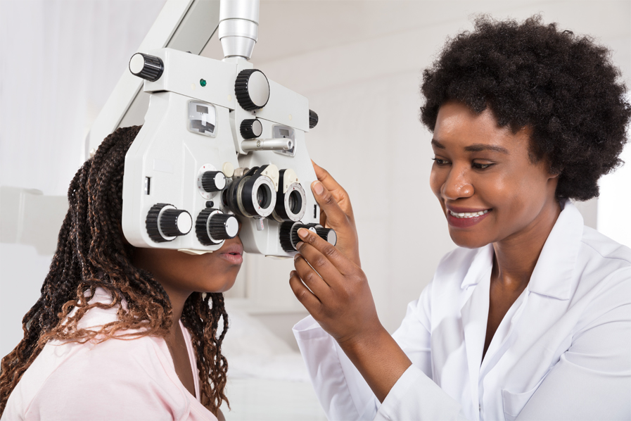
REFRACTION
Determining refractive error with a phoropter and retinoscope
Refraction is conducted to determine the appropriate lens power needed to compensate for any refractive error (nearsightedness, farsightedness, or astigmatism). Using an instrument called a phoropter, your optometrist places a series of lenses in front of your eyes and measures how they focus light using a hand- held lighted instrument called a retinoscope. The optometrist may choose to use an automated instrument that automatically evaluates the focusing power of the eye. The power is then refined by patient's responses to determine the lenses that allow the clearest vision.
This testing may be done without the use of eye drops to determine how the eyes respond under normal seeing conditions. In some cases, such as for patients who can't respond verbally or when some of the eyes focusing power may be hidden, eye drops are used. The drops temporarily keep the eyes from changing focus while testing is done.
EYE FOCUSING, EYE TEAMING, AND EYE MOVEMENT TESTING
Assessment of accommodation, ocular motility and binocular vision determines how well the eyes focus, move and work together. In order to obtain a clear, single image of what is being viewed, the eyes must effectively change focus, move and work in unison. This testing will look for problems that keep your eyes from focusing effectively or make using both eyes together difficult.
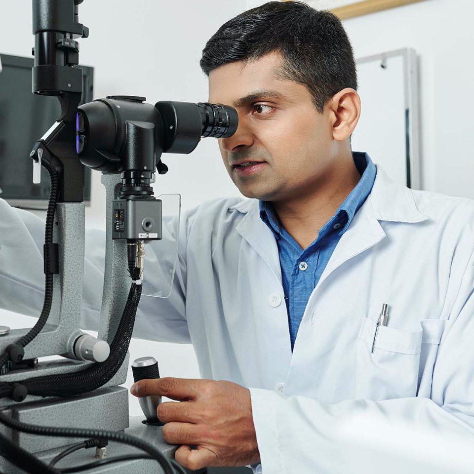
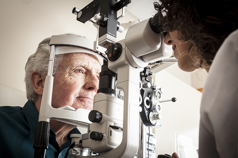
EYE HEALTH EVALUATION
Tonometry measures eye pressure. Elevated pressure in the eye signals an increased risk for glaucoma.
External examination of the eye includes evaluation of the cornea, eyelids, conjunctiva and surrounding eye tissue using bright light and magnification. Evaluation of the lens, retina and posterior section of the eye may be done through a dilated pupil to provide a better view of the internal structures of the eye. Measurement of pressure within the eye (tonometry) is performed. Normal eye pressures range from 10 to 21 millimetres of mercury (mm Hg), averaging about 14 to 16 mm Hg. Anyone with eye pressure greater than 22 mm Hg is at an increased risk of developing glaucoma, although many people with normal pressure also develop glaucoma.
SUPPLEMENTAL TESTING
Additional testing may be needed based on the results of the previous tests to confirm or rule out possible problems, to clarify uncertain findings, or to provide a more in-depth assessment.
At the completion of the examination, your optometrist will assess and evaluate the results of the testing to determine a diagnosis and develop a treatment plan. He or she will discuss with you the nature of any visual or eye health problems found and explain available treatment options. In some cases, referral for consultation with, or treatment by, another optometrist or other health care provider may be indicated.
If you have questions regarding any eye or vision conditions diagnosed, or treatment recommended, don't hesitate to ask for additional information or explanation from your SAOA optometrist.
A comprehensive eye exam is not just a vision screening. Remember the eye test with the tiny letters you took at school or to renew your driver’s license? That’s a vision screening.
A vision screening isn't enough
If you’re wondering what the difference is between a vision screening test (the kind you received in school) and a comprehensive eye exam, a vision screening only indicates a potential need for further evaluation. Even the most sophisticated vision screening tools, administered by the most highly trained screeners, can miss children with eye or vision disorders.
Screening will only tell you if you would benefit from spectacles and will not tell if your eyes are healthy. Not being able to see well, is the most common sign of eye problems including eye disease, that can cause blindness. Therefore, it is always necessary for an optometrist to examine your eyes thoroughly to make a diagnosis before prescribing a treatment.
Periodic eye and vision examinations are an important part of preventive health care. Many eye and vision problems have no obvious signs or symptoms. As a result, individuals are often unaware that problems exist. Early diagnosis and treatment of eye and vision problems are important for maintaining good vision and eye health, and when possible, preventing vision loss.
An eye examination takes a while so you should expect to be in the chair for at least 30 minutes and maybe more. For many people the news will be that they have great eye health. Approximately 75 percent of people examined need to be prescribed spectacles or contact lenses for refractive error.
A comprehensive adult eye and vision examination may include but is not limited to the tests mentioned under ’what to expect from an eye exam?’ Individual patient signs and symptoms, along with the professional judgment of the optometrist, may significantly influence the testing done.
There's no app for that
You may have seen apps advertised that can ‘replace going to the eye care professional’ However, online vision tests attempt to replace one element of an eye examination, the refraction, to yield a prescription for spectacles or contact lenses. It is not unlike taking a blood pressure reading at a kiosk and expecting a prescription – the reading does not provide sufficient information to determine a patient's needed course of therapy. The refraction performed by online vision tests is only one of many tests performed during an eye examination, and taken by itself, does not provide sufficient information regarding the treatment of a patient, including the prescription of glasses or contact lenses.
Are online vision tests accurate?
Online vision tests may give inaccurate or misleading information, and can give patients a false sense of security. In fact, patients may delay essential, sight-saving treatment. Comprehensive eye exams with your optometrist are one of the most important, preventive ways to preserve vision, and the only way to accurately assess eye health, diagnose an eye disorder or disease, and determine the need for corrective lenses.
Children Vision
SAOA Optometrists, through their clinical education, training and experience have the means to provide effective primary eye and vision services to children. Vision care and regular eye examinations are essential for children, the earlier the better. According to the South African Optometric Association (SAOA) early detection of visual problems followed by early intervention in vision care is crucial to achieve normal vision in infants and young children with visual anomalies.
Signs to watch out for in children
Vision Problems in children
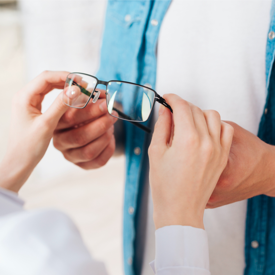
If interested in wearing contact lenses, get off to the right start by seeing an optometrist. An optometrist will provide a thorough eye examination and an evaluation of suitability for contact lens wear.
What is the best contact lens care system?
Contact lens care systems are highly important to the success of wearing contact lenses and also the reduction of risk for infections. A contact lens care system has been carefully selected by your optometrist in order to be compatible with the lens materials as well as the way you wear your lenses eg daily disposable, monthly disposable, extended wear or specialized lenses.
There are many different types of contact lens care systems, but the basic two types are multipurpose care solutions and hydrogen peroxide care solutions. For gas permeable (GP) lenses, your solutions may contain either a one-step (multipurpose) solution or a two-step (cleaning and soaking) solution or you may also use a hydrogen peroxide solution. These types of solutions all have different recommendations for use depending on the manufacturer. Consult the package insert or your optometrist for the best practices regarding the use of your lens care system.
Lens materials and contact lens solutions can interact, which can also affect the disinfection process. Generic (or store brand) contact lens solutions may have been formulated for older lens materials; new lens materials have different chemical compositions and may not be compatible with generic solutions. If you wish to change your contact lens care system, contact your optometrist first to make sure it is safe for the particular type of contact lenses that you wear.
Do I need to rub my contact lenses when I take them out?
Yes, if you are using a multipurpose solution with soft contact lenses (there are currently no recommended “no-rub” multipurpose contact lens solution regimens). Rubbing the contact lens for between two and 20 seconds, depending on your contact lens care solution, removes deposits and micro-organisms and reduces complications. You follow the rubbing step with a thorough rinse with solution for the time specified by the manufacturer (usually between five and 10 seconds). Recent evidence conclusively demonstrated that rubbing and rinsing the lens after wear provides the safest lens wear for all contact lenses and care systems currently on the market. Some hydrogen peroxide systems contain a rinse step prior to soaking the lenses overnight. Gas permeable (GP) lenses also contain a rub step with hydrogen peroxide systems prior to this rinse step. If you are unsure how to care for your lenses, please refer to the manufacturer’s instructions provided with your care system or contact your optometrist.
Are my solutions good past the expiration date?
Contact lens solutions should never be used after the expiration date listed on the packaging. The United States Food and Drug Administration (US FDA) has also recommended that solution manufacturers begin labelling solution bottles with a discard date, in addition to the usual expiration date. The discard date is the date the solution should be thrown out after opening (typically within 90 days but varies depending on the manufacturer and solution type). Refer to the solution’s package insert for directions and the discard dates and be sure to ask your optometrist if you are unsure of how to use your prescribed care solutions. It should be noted that preservative-free contact lens solutions (such as unit-dose single vial saline) should be discarded within 24 hours after opening. Saline that is packaged in aerosol containers typically has a longer discard date. Refer to the solution’s package insert for more information or contact your optometrist.
Can I use any lubricants or red-eye drops with contact lenses?
Any eye drops not approved for contact lens wear can cause damage to both the contact lens and the eye. Stay away from drops that claim to “get the red out,” as they typically contain chemicals that may be detrimental to your long-term eye health. Preservative-free eye drops, in general, are very safe to use with contact lenses. Eye drops that contain preservatives can have a toxic effect on the eye and should be avoided. Consult an optometrist about which drops are best for your eyes and contact lens materials.
I only wear my lenses occasionally. How should I store them in the meantime?
Care systems for contact lenses are all very different, and it is important to note how long contact lenses can be safely stored in solution long term before the solution needs to be replaced. Some solutions are only good for 24 hours of storage while others can store lenses for up to one month.
Some soft contact lens multipurpose solutions allow for long-term storage of up to one month in a tightly closed contact lens case. For GP lenses, some GP lens solutions also allow for one month of long-term storage. Some hydrogen peroxide-based systems allow for seven days of long-term storage before the lenses need to be re-disinfected but others are as short as 24 hours of storage time. The night before you are ready to wear your lenses again, it is a good idea to inspect the lens and then re-clean and disinfect it according to the solution manufacturer’s instructions. Check the manufacturer’s recommended storage times on the package insert or contact your optometrist for further guidance if needed.
If you only wear lenses on occasion, a daily disposable contact lens may be an option for you. Daily disposable lenses are great for part-time wearers to keep costs down and ensure a safe contact lens wear experience. You don’t need to worry about how long your lenses have been sitting in solution or whether your solution is past its discard date. Daily disposable lenses are also highly portable and convenient. In addition, daily disposable lens wearers experience fewer contact lens complications like infections when compared to other lens replacement schedules.
What are the proper steps to follow each time I remove my contact lens from my eye?
Contact lenses are all taken care of differently; this is an example of how most soft contact lenses are cared for after removal in conjunction with a multipurpose solution (rub and rinse times may vary depending on solution manufacturer). Hydrogen peroxide care systems are different from below. Keep in mind that daily disposable contact lenses are removed each night and thrown away, so no cleaning is required.
I dropped the contact lens on the floor, but I don’t have time to run an entire disinfection cycle with my solution. What do I do?
It is not enough simply to rinse off the contaminants and dust particles from the floor. Bathroom floors and other such surfaces may harbour significantly high numbers of microorganisms. While the tear film and corneal surface provide a remarkable barrier to infection, one never knows when the barrier may break down.
The only sure way to guarantee safety is to apply a new contact lens. This can be easily accomplished if you are wearing disposable soft contact lenses. The next best thing is to wear your back-up glasses while you clean the lens through the entire disinfection cycle as recommended by your care system's manufacturer. Every contact lens wearer should have a pair of glasses to wear as needed in cases like these when their lenses cannot be worn.
Is it okay to use tap water to insert or store my lenses?
No. Tap water contains micro-organisms which can lead to serious eye infections and loss of vision. One of the more well-known water-borne infections is caused by Acanthamoeba, a microscopic, free-living amoeba (single-celled organism). Acanthamoeba can cause an infection of the cornea, the transparent outer covering of the eye where a contact lens sits. These organisms are very common in nature and can be found in bodies of water (for example, lakes and oceans), hence the recommendation to avoid the use of water with lenses.
You should never use tap water in any area of your lens care, including rinsing the lenses and the lens case. Do not attempt to make your own homemade saline or contact lens solutions. Also, make sure your hands are completely dry before handling your lenses.
Should I be concerned about wearing my contact lenses on an airplane?
Commercial airline cabins expose passengers to reduced atmospheric pressure, reduced oxygen availability, reduced humidity, and dry air. These conditions can lead to discomfort with contact lens wear, especially on flights lasting longer than three hours. Instillation of lubricating eye drops approved for use with contact lenses may help relieve some eye dryness during your flight. Keep in mind that the South African Civil Aviation Authority limits the size of any liquid container carried onboard an airplane to a 100 ml bottle or less unless the liquids are medically necessary. Contact lens solution is considered a medically necessary liquid, so full-size bottles must be declared separately at the entry to the security checkpoint. The solution is then subject to further screening. Solution manufacturers do tend to sell 60ml travel size containers of solution for those still wary of restrictions. In the absence of a smaller manufacturer-supplied container of solution, do not attempt to transfer contact lens solution to a smaller container. This allows for contamination of the solution during transfer, which can lead to a serious eye infection.
Is it safe to wear my contact lenses in a bathtub or hot tub?
It is recommended not to wear contact lenses while in a bathtub or hot tub. The South African Optometric Association has recommended that contact lenses not be exposed to any form of water. Although rare, a sight-threatening eye complication known as Acanthamoeba keratitis is caused by an organism present in all forms of impure water (e.g. swimming pools, tap water, saunas, wells, and showers). Acanthamoeba and certain forms of bacteria present in water can become attached to contact lenses, resulting in potential infection.
If lenses are worn while in the bathtub or hot tub, care should be taken to avoid water being splashed into the eyes. Recent studies have recommended the use of tight-fitting swim goggles to limit eye exposure to water while swimming. If lenses are accidentally exposed to water, instil a lubricating drop to help loosen the lens on the eye then remove the lens with clean, dry hands. Next, clean and disinfect the lens before re-inserting, or discard the lens. Never sleep in a lens that has been exposed to water without first cleaning and disinfecting it. If lenses were removed prior to getting in a bathtub or hot tub, they must be properly cleaned and disinfected before being re-inserted.
Contact lens do’s
Contact lens don'ts
Signs of potential problems
It is generally not difficult to wear contact lenses. Following your optometrist’s advice and regular follow-up care will prevent most problems.
However, here is a list of some signs that things may not be going well. If you experience any of these, contact your optometrist as soon as possible.
Contact lenses and cosmetics
You can wear contacts and cosmetics safely and comfortably together by following these helpful tips:
There's no substitute for the quality of life good vision offers. Adding certain nutrients to your daily diet—either through foods or supplements—can help preserve your vision. Researchers have linked eye-friendly nutrients, such as lutein and zeaxanthin, vitamin C, vitamin E, and zinc, to a reduction in the risk of certain eye diseases.
Lutein & Zeaxanthin
Lutein and zeaxanthin are important nutrients found in green leafy vegetables, as well as other foods, such as eggs. Many studies show that lutein and zeaxanthin reduce the risk of chronic eye diseases, including age-related macular degeneration and cataracts.
Vitamin C
Vitamin C (ascorbic acid) is an antioxidant found in fruits and vegetables. Scientific evidence suggests vitamin C lowers the risk of developing cataracts. Also, when taken in combination with other essential nutrients, it can slow the progression of age-related macular degeneration and visual acuity loss.
Vitamin E
Vitamin E is a powerful antioxidant found in nuts, fortified cereals, and sweet potatoes. Research indicates it protects cells in the eyes from unstable molecules called free radicals, which break down healthy tissue.
Essential fatty acids
Fats are a necessary part of the human diet. They maintain the integrity of the nervous system, fuel cells and boost the immune system. Research shows omega-3 fatty acids are important for proper visual development and retinal function.
Zinc
Zinc is an essential trace mineral or "helper molecule." It plays a vital role in bringing vitamin A from the liver to the retina in order to produce melanin, a protective pigment in the eyes. Zinc is highly concentrated in the eye, mostly in the retina and choroid, the vascular tissue layer lying under the retina.
Emerging research
In the last 20 years, eye health research has linked diet and nutrition with a decreased risk of age-related macular degeneration (AMD). A major clinical study of older adults concluded that taking an antioxidant vitamin or mineral supplement significantly reduced the risk of advanced AMD progression in some people. Additionally, today there is significant evidence that vitamin D plays a role in preventing AMD.
AREDS made it clear
The Age-Related Eye Disease Study (AREDS) was a major clinical trial sponsored by the National Eye Institute. It enrolled 3640 subjects, age 55 to 80, and was released in October 2001. This landmark study provided evidence that nutritional intervention in the form of supplements could delay the progression of AMD. The study concluded that taking an antioxidant vitamin or mineral supplement reduced the risk of advanced AMD progression by about 25 percent and showed a 19 percent reduction in visual acuity loss in some of the subjects.
Nutrition
Many optometrists are expanding their traditional roles to include other areas that affect eye health, such as nutritional education. Research has shown that certain nutrients can delay the development and progression of cataracts and AMD. These are two of the leading causes of blindness and visual impairment for millions of aging South Africans. Nutrition may be particularly important in treating AMD. Currently, treatment options after diagnosis of this condition are limited. Also, people who suffer from dry eye, an increasing problem due to the visual demands of digital devices, can often benefit from nutritional supplements.
Hyperopia
This is one form of refractive error, in which distant objects are usually seen clearly, but close ones do not come into proper focus.
How does Hyperopia develop?
Hyperopia occurs if your eyeball is too short or the cornea has too little curvature. Light entering your eye is not focused correctly. As a result, near objects cannot be brought into sharp and clearly focused images. Hereditary factors often control the growth and development of the eye. However, environmental factors may also contribute to the development of farsightedness.
What causes Hyperopia?
People with hyperopia involuntarily exert extra effort to maintain clear distance vision and even greater effort to see clearly at close range. This extra effort can cause fatigue, tension and discomfort. If the crystalline lens of the eye cannot bring the object being viewed into focus, blurred vision occurs.
What are the signs and symptoms of Hyperopia?
Difficulty in concentrating and maintaining a clear focus on near objects,
How is Hyperopia diagnosed?
Common vision screenings, often done in schools, are generally ineffective in detecting hyperopia. This is because these individuals can identify the letters on an eye chart with little difficulty. However, a comprehensive optometric examination is preferred in diagnosing hyperopia.
How is Hyperopia corrected?
In mild cases of hyperopia, your eyes may be able to compensate without corrective lenses. While in other cases, there is a need for the optometrist to prescribe spectacles or contact lenses to optically correct hyperopia. For more information visit your nearest SAOA Optometrist.
Myopia
This is also known by many as short or near sightedness. This results in distance objects appearing blurry while close objects appear clearly. This is a common type of refractive error and usually corrected by spectacles or contact lenses. There are however, special cases that need laser eye surgery.
How does myopia develop? Myopia develops when the images in the eyes focus in front of the retina instead of on the retina. This results in blurred vision. It is caused when the eyeball becomes too long and prevents incoming light from focusing directly on the retina. It may also be caused by an abnormal shape of the cornea or lens.
What causes myopia?
Myopia can affect both children and adults. Myopia is often diagnosed in children between 8 and 12 years of age and may worsen during the teen years. Little change may occur between ages 20 to 40, but sometimes myopia may worsen with age. People with a positive family history of myopia may be more likely to get the condition.
Myopia, in children, has been linked to excessive near activities like the use of digital devices and smartphones.
What are the signs and symptoms of myopia?
Some of the signs and symptoms of myopia include:
How is myopia diagnosed?
Regular comprehensive eye examinations are advised. It is during the eye examination that an Optometrist can diagnose and advise on what treatment is best suited for your eyes.
How is myopia corrected?
Myopia can be corrected with the use of spectacles, contact lenses, or surgery. Spectacles are the simplest and safest way to correct myopia. Your Optometrist can prescribe lenses that will correct the problem and help you to see well. Contact Lenses work by being in contact with the eye, thus providing a refractive surface for light rays entering the eye, enabling clearer and better focus. In many cases, contact lenses provide clearer vision, a wider field of vision, and greater comfort. They are a safe and effective option if fitted and used properly. However, contact lenses are not right for everyone. For more information, visit your nearest SAOA Optometrist.
Myopia Control
‘Myopia control’ is the term used to describe specific treatments to slow the progression of nearsightedness in children. Myopia control measures are prescribed by the SAOA optometrist.
There are four primary categories of myopia control treatments:
Myopia control is important because it may help reduce the risk of vision-threatening complications associated with high myopia later on in life. These include glaucoma, cataracts, retinal detachment and even blindness.
What is astigmatism?
Astigmatism is a common vision problem caused by an error in the shape of the cornea. With astigmatism, the lens of the eye or the cornea, which is the front surface of the eye, has an irregular curve.
This can change the way light passes, or refracts, to your retina. This causes blurry, fuzzy, or distorted vision.
Farsightedness(hyperopia) and Nearsightedness(myopia) are two other types of problems with the way light passes to your retina.
Farsightedness or hyperopia causes patients to have more problems at near. Nearsightedness or myopia, causes patients to have more problems at far distances.
What are the types of astigmatism?
The two main types of astigmatism are corneal and lenticular. A corneal astigmatism happens when your cornea is more oval then spherical. A lenticular astigmatism happens when your lens has the same oval shape. Corneas should have an ideal spherical shape.
What causes astigmatism?
It’s not known what causes astigmatism, but genetics is a big factor. It’s often present at birth, but it may develop later in life. It may also occur as a result of an injury to the eye or after eye surgery. In teenagers, it can often develop as a result of eye rubbing due to allergies. High astigmatism caused by eye rubbing, can lead to a condition called Keratoconus. Eye rubbing should therefore be avoided Astigmatism often occurs with nearsightedness or farsightedness.
Who is at risk for astigmatism?
Astigmatism can occur in children and adults. Your risk of developing astigmatism may be higher if you have any of the following:
What are the symptoms of astigmatism?
The symptoms of astigmatism may differ in each person. Some people don’t have any symptoms at all. The symptoms of astigmatism include:
Visit an Optometrist if you have symptoms of astigmatism. Some symptoms may also be due to other health or vision problems. The Optometrist will then refer the patient for further tests.
How is astigmatism diagnosed?
An optometrist diagnoses astigmatism through a comprehensive eye health and vision examination. An optometrist is a professional who diagnoses vision problems and eye diseases. An ophthalmologist is a doctor who provides medical and surgical treatment of vision problems and eye diseases. There are several tests an optometrist may use during your eye examination to diagnose astigmatism.
What is Presbyopia?
Presbyopia is a vision condition in which the crystalline lens of your eye loses its flexibility, which makes it difficult for you to focus on close objects.
Presbyopia may seem to occur suddenly, but the actual loss of flexibility takes place over a number of years. Presbyopia usually becomes noticeable in the early to mid-40s. Presbyopia is a natural part of the aging process of the eye. It is not a disease, and it cannot be prevented.
What are the signs and symptoms of Presbyopia?
Some signs of presbyopia include the:
Optical solutions include:
How is presbyopia corrected?
A Comprehensive optometric examination will include testing for presbyopia.
To help you compensate for presbyopia, your SAOA optometrist may prescribe any of the above-mentioned corrections. Presbyopia can complicate other common vision conditions like myopia, hyperopia and astigmatism, your optometrist will determine the specific lenses to allow you to see clearly and comfortably.
You may need to wear your glasses for close work like reading, but you may also find that wearing them all the time is more convenient and beneficial for your vision needs. The effects of presbyopia continue to change the ability of the crystalline lens to focus properly, meaning that regular changes in your spectacles may be necessary to maintain clear and comfortable vision. For more information visit your nearest SAOA Optometrist.
Refractive errors are vision related problems. These are the common reasons for reduced levels of eyesight (visual acuity). What are refractive errors? In refractive errors, the shape of the eye prevents light from focusing on the retina. The length of the eyeball (longer or shorter), changes in the shape of the cornea, or aging of the lens can cause refractive errors.
There are four types of refractive error:
Glaucoma
What is diabetes?
Diabetes is described as a group of metabolic diseases in which the person has high blood glucose (blood sugar), either because insulin production is inadequate, or because the body's cells do not respond properly to insulin, or both. The effect of diabetes on the eye is called diabetic retinopathy. Diabetic retinopathy is a common complication of diabetes, it may not have any symptoms or may not affect sight in the early stages but, as the condition progresses, eventually the sight will be affected. When the condition is caught early, treatment is effective at reducing or preventing damage to sight.
People with high blood sugar may experience some of these symptoms:
Types of Diabetes
The following increases your chance of developing diabetes
What do you need to do?
Measuring the glucose level in blood. The first stage would be to make some lifestyle change, such as:
Getting better control over your blood sugar, cholesterol, and blood pressure levels can help reduce the risk of kidney disease, eye disease, nervous system disease, heart attack, and stroke. Keeping an ideal body weight and an active lifestyle may prevent type 2 diabetes. Type 1 diabetes cannot be prevented.
How do you treat it?
Non-insulin dependent diabetes (Type 2) develops slowly; some people with high blood sugar have no symptoms at the time of the diagnosis. This making it extremely important for people with diabetes to have annual dilated eye examinations, as it can help in early detection of diabetes changes in the eye. The longer you have the diabetes the higher the risk of developing diabetic eye disease. Many problems can be treated with much greater success when caught early. There are many ways of treating diabetic retinopathy, which depends on the stage of the disease and the specific problem that requires attention. The goal of the treatment is to arrest the progression of the disease. A Multidisciplinary approach is ideally used with diabetes, such as Optometrist, Ophthalmologist, Podiatrist and Endocrinologist. For more information visit your nearest SAOA Optometrist.
Useful Links
References
Often referred to casually as “pink eye”, conjunctivitis is the swelling or inflammation of the conjunctiva, the thin, transparent layer of tissue that lines the inner surface of the eyelid and covers the white part of the eye. Causes may or may not be infectious.
Causes & risk factors
There are three main types of conjunctivitis: allergic, infectious and chemical. The cause of conjunctivitis varies depending on the type.
Allergic conjunctivitis
Infectious conjunctivitis
Chemical conjunctivitis
Chemical Conjunctivitis can be caused by irritants like air pollution, chlorine in swimming pools, and exposure to noxious chemicals.
Symptoms
Symptoms vary with the causes discussed above. Allergic symptoms include clear, watery discharge along with mild redness. Itching, sometimes severe, may or may not occur. With bacterial infections, there is typically minimal pain but a possibly dramatic appearance with moderate redness and almost always a yellow/green discharge, sometimes extreme. This discharge can also make the eyelids red and swollen and can attach itself to the eyelashes for a crusty appearance.
Bacterial infections can be more severe in patients that wear contact lenses. There is also a risk of a bacterial corneal ulcer developing in contact lens wearers which would include severe pain and light sensitivity. Viral infections can also cause moderate redness and are usually painful. The pain is typically a sandy, gritty feel like something may be in the eye. There can also be a moderate to severe light sensitivity.
Diagnosis
Conjunctivitis can be diagnosed through a comprehensive eye examination. Testing, with special emphasis on the conjunctiva and surrounding tissues, may include:
Using the information obtained from these tests, an optometrist can determine if you have conjunctivitis and provide further referral for care
Treatment
Treating conjunctivitis has three main goals:
The appropriate treatment for conjunctivitis depends on its cause.
Allergic conjunctivitis
The first step is to remove or avoid the irritant, if possible. Cool compresses and artificial tears sometimes relieve discomfort in mild cases. In more severe cases, nonsteroidal anti-inflammatory medications and antihistamines may be prescribed. People with persistent allergic conjunctivitis may also require topical steroid eye drops. Oral antihistamines may also be prescribed.
Infectious conjunctivitis
This type of conjunctivitis is usually treated with antibiotic eye drops or ointments. Bacterial conjunctivitis may improve after three or four days of treatment, but patients need to take the entire course of antibiotics to prevent a recurrence. Viral conjunctivitis. No drops or ointments can treat viral conjunctivitis. Antibiotics will not cure a viral infection. Like a common cold, the virus has to run its course, which may take up to two or three weeks. Symptoms can often be relieved with cool compresses and artificial tear solutions. For the worst cases, topical steroid drops may be prescribed to reduce the discomfort from inflammation. However, these drops will not shorten the infection. The viral infection Epidemic Keratoconjunctivitis (EKC) is very contagious and is the red-eye most associated with the term “pink eye”.
Chemical conjunctivitis
Careful flushing of the eyes with saline is a standard treatment for chemical conjunctivitis. People with chemical conjunctivitis also may need to use topical steroids. Severe chemical injuries, particularly alkali burns, are medical emergencies and can lead to scarring, damage to the eye or the sight, or even loss of the eye. If a chemical spills in your eye, flush the eye for several minutes with a lot of water before seeing your medical provider.
Contact lens wearers may need to temporarily stop wearing their lenses while the condition is active. If conjunctivitis is due to wearing contact lenses, an optometrist may recommend switching to a different type of contact lens or disinfection solution. An optometrist might need to change the contact lens prescription to a lens that is replaced more frequently. This can help prevent conjunctivitis from recurring.
Practicing good hygiene is the best way to control the spread of conjunctivitis. Once an infection has been diagnosed, follow these steps:
Soothe the discomfort of viral or bacterial conjunctivitis by applying warm compresses to your affected eye or eyes. To make a compress, soak a clean cloth in warm water and wring it out before applying it gently to your closed eyelids. For allergic conjunctivitis, avoid rubbing the eyes. Instead of warm compresses, use cool compresses to soothe your eyes. Over-the-counter eye drops might also help. Antihistamine eye drops can alleviate the symptoms, and lubricating eye drops can rinse the allergen off the surface of the eye. See your optometrist if you think you have conjunctivitis. He or she can diagnose the cause and prescribe the proper treatment or refer to the ophthalmologist
Prevention
With so many causes, there is no one preventive measure. Early diagnosis and treatment will help prevent the condition from becoming worse. Avoiding allergy triggers as much as possible also helps. Frequent hand washing and keeping hands away from eyes also can make a difference, even when no problems are present.
Dry eye is a condition in which a person doesn't have enough quality tears to lubricate and nourish the eye. Tears are necessary for maintaining the health of the front surface of the eye and for providing clear vision.
Dry eye is a common and often chronic problem, particularly in older adults. With each blink of the eyelids, tears spread across the front surface of the eye, known as the cornea. Tears provide lubrication, reduce the risk of eye infection, wash away foreign matter in the eye and keep the surface of the eyes smooth and clear. Excess tears in the eyes flow into small drainage ducts in the inner corners of the eyelids, which drain into the back of the nose.
Causes & risk factors
Dry eyes can occur when tear production and drainage are not in balance. People with dry eyes either do not produce enough tears or their tears are of a poor quality:
Reasons for developing dry eyes
Advanced dry eyes may damage the front surface of the eye and impair vision.
Symptoms
People with dry eyes may experience irritated, gritty, scratchy or burning eyes; a feeling of something in their eyes; excess watering; and blurred vision. Symptoms include:
Diagnosis
Dry eyes can be diagnosed through a comprehensive eye examination. Testing with emphasis on the evaluation of the quantity and quality of tears produced by the eyes may include:
With the information obtained from testing, an optometrist can determine if you have dry eyes and advise you on treatment options.
Treatment
Treatments for dry eyes aim to restore or maintain the normal amount of tears in the eye to minimize dryness and related discomfort and to maintain eye health. Dry eyes can be a chronic condition, but an optometrist can prescribe treatment to keep your eyes healthy and comfortable and to prevent your vision from being affected. The primary approaches used to manage and treat dry eyes include adding tears using over-the-counter artificial tear solutions, conserving tears, increasing tear production, and treating the inflammation of the eyelids or eye surface that contributes to the dry eyes.
With the information obtained from testing, an optometrist can determine if you have dry eyes and advise you on treatment options.
Prevention
You can take the following steps to reduce symptoms of dry eyes:
With the information obtained from testing, an optometrist can determine if you have dry eyes and advise you on treatment options.
Keratoconus is a vision disorder that occurs when the normally round cornea (the front part of the eye) becomes thin and irregular (cone) shaped. This abnormal shape prevents the light entering the eye from being focused correctly on the retina and causes distortion of vision.
Causes & risk factors
Symptoms
In its earliest stages, keratoconus causes slight blurring and distortion of vision and increased sensitivity to glare and light. These symptoms usually appear in the late teens or early 20s. Keratoconus may progress for 10-20 years and then slow in its progression. Each eye may be affected differently. As keratoconus progresses, the cornea bulges more and vision may become more distorted. In a small number of cases, the cornea will swell and cause a sudden and significant decrease in vision. The swelling occurs when the strain of the cornea's protruding cone-like shape causes a tiny crack to develop. The swelling may last for weeks or months as the crack heals and is gradually replaced by scar tissue. If this sudden swelling does occur, your doctor can prescribe eyedrops for temporary relief.
Diagnosis
See an optometrist regularly for corneal curvature measurements and tests for irregular astigmatism.
Treatment
Spectacles or soft contact lenses may be used to correct the mild near-sightedness and astigmatism that is caused by the early stages of keratoconus. Corneal cross-linking surgery is indicated early after diagnosis to stabilize the structure of the cornea and slow progression. As the disorder progresses and the cornea continues to thin and change shape, rigid gas permeable lenses and scleral lenses, can be prescribed to correct vision adequately. In most cases, this is sufficient. The contact lenses must be carefully fitted, and frequent check-ups and lens changes may be needed to achieve and maintain good vision. In a few cases, a corneal transplant is necessary. However, even after a corneal transplant, spectacles or contact lenses are often still needed to correct vision.
The Corneal cross-linking procedure halts or slows progression and is indicated early after diagnosis. It can be done later, if in the course of the disease, there is still an increase in the spectacle prescription. Intacs, which are intracorneal removable inserts can stretch the cornea to stabilize astigmatism and near-sighted refractive errors. Once the progression has stabilized from cross-linking, an implantable Collamer lens surgery can correct higher prescriptions so that spectacles or conventional soft contact lenses can be utilized.
An optometrist is a healthcare professional who provides primary vision care. An optometrist has a university degree and this qualification enables the practicing of optometry, which includes performing comprehensive vision and eye health examinations, prescribing & dispensing corrective lenses, detecting certain eye abnormalities, and prescribing medications for certain eye diseases.
Optometrists are trained to examine eyes to detect defects in vision, signs of injury, ocular diseases or abnormality and problems with general health, such as high blood pressure or diabetes. They make a health assessment, offer clinical advice, carry out vision therapy, prescribe spectacles, contact lenses & therapeutic pharmaceuticals when necessary. Patients are referred for further treatment, when required. All optometrists practicing in South Africa must be registered with the profession’s regulatory body, the Health Professions Council of South Africa (HPCSA).
Qualified optometrists are encouraged to belong to the South African Optometric Association (SAOA). The SAOA guides the profession & advises the public on all optometry related matters.
A optician, or dispensing optician, is a technical practitioner who designs, fits and dispenses spectacle and contact lenses for the correction of a person's vision. Opticians determine the specifications of various ophthalmic appliances that will give the necessary correction to a person's eyesight.
An ophthalmologist — Eye M.D. — is a medical doctor, who has continued training, and now specializes in eye and vision care. Ophthalmologists differ from optometrists and opticians in their levels of training and in what they can diagnose and treat. As a medical doctor who has completed a university degree and at least four years of additional medical training, an ophthalmologist is licensed to practice medicine and surgery. An ophthalmologist diagnoses and treats all eye diseases, performs eye surgery and most often refers to optometry to fit spectacles and contact lenses to correct vision problems. Many ophthalmologists are also involved in scientific research on the causes and cures for eye diseases and vision disorders.
Ophthalmologists are surgical and medical specialists able to perform operations on eyes. Medically qualified, they mainly work in eye hospitals and hospital eye clinics. Visit the Ophthalmological Society of South Africa (OSSA) to find out more.
Orthoptics is a profession allied to the eye care professions of Optometry and Ophthalmology. Orthoptists are experts in diagnosing and treating defects in eye movements and eye alignment. Patients with squints, for example, have problems with how their eyes work together which leads to binocular vision difficulties.
ABOUT US
FIND AN OPTOM
CONTACT US